
Relationship of one's FAZ section into the FA having those who work in the shallow layer-on OCTA (a) and you will dentro de-face October photo (b) and those in the strong layer-on OCTA (c) and you may dentro de-deal with Oct images (d). (elizabeth,f) The partnership between the FAZ portion regarding the OCTA and you may durante-face Oct photo regarding the superficial otherwise strong layer. ICC = intraclass coefficient.
Particular microaneurysms are very well-laid out regarding the en-deal with October images by yourself (green arrowheads) or even in each other (red arrowheads)
We also investigated the microaneurysms in the OCTA and en-face OCT images that corresponded to the hyperfluorescent dots in the early and late phases. The decorrelation signals in the OCTA images showed a few morphologic patterns, i.e., fusiform, saccular, curved and rarely coiled, compared to the homogeneous dot-like signals in the FA images (Figs 5d,g and 6d,g). Microaneurysms appeared in the en-face OCT images as highly reflective round, oval, or sometimes ringed lesions in DR, which agreed with recent studies about B-scan images (Figs 5e,h and 6e,h) 21,22,23 . We counted microaneurysms in the FA, OCTA and en-face OCT images, which had good intergrader compatibility (ICC = 0.956, 0.939 and 0.917 in the FA, OCTA and en-face OCT images, respectively) and compared microaneurysms in the OCTA or en-face OCT images to those in the FA images. Only 41.0 ± 16.1% of the microaneurysms in the FA images showed such lesions in the OCTA images in all 53 eyes. En-face OCT images delineated 40.1 ± 18.6% of the microaneurysms in the FA images. The numbers of microaneurysms in the OCTA and en-face OCT images were associated significantly (? = 0.821, P < 0.001and ? = 0.782, P < 0.001, respectively) with the number in the FA images (Fig. 7a,b). Further studies showed that 13.9 ± 16.4% of microaneurysms were seen in both the OCTA and en-face OCT images. Either OCTA or en-face OCT images detected 67.3 ± 18.3% of the microaneurysms in the FA images and the numbers of microaneurysms in either image were significantly correlated with that in the FA images (? = 0.899, P < 0.001) (Fig. 7c).
(b,c) Early- and you will later-phase FA photo of the areas equal to the latest black colored square regarding the color fundus image (a) reveal microaneurysms as the hyperfluorescent dots (arrow and you will arrowheads). Some microaneurysms are seen from the OCTA pictures by yourself (red-colored arrowhead) or in the en-deal with October photos alone (environmentally friendly arrowhead) or even in one another (red-colored arrowhead). Someone else aren't observed in either the new OCTA otherwise en-deal with Oct visualize (arrow). (d,g) OCTA photos. (elizabeth,h) En-deal with October images. (f,i) Combined pictures out-of OCTA (red) and durante-deal with October (gray). Superficial (d–f) or strong (g–i) level.
The early- or later-phase (b,c) FA pictures of the areas corresponding to the new black rectangular in the colour fundus pic (a). Specific microaneurysms have emerged regarding OCTA pictures by yourself (red arrowheads) and possess some morphologies, i.elizabeth., fusiform, saccular and coiled. Anybody else aren't depicted either in the latest OCTA or dentro de-face October visualize (arrow). Microaneurysms in a choice of the new low (d–f) or deep (g–i) level are occasionally right beside cystoid rooms. (d,g) OCTA photographs. (elizabeth,h) En-deal with October pictures. (f,i) Merged pictures out of OCTA (red) and en-deal with October (gray).
Comparison regarding microaneurysms to the fluorescein angiography (FA) with optical coherence tomography angiography (OCTA) (a), en-deal with optical coherence tomography (OCT) (b), otherwise OCTA and/otherwise dentro de-face October photos (c) in the diabetic retinopathy.
Talk
Capillary nonperfusion has actually a life threatening impact on visual impairment and you may leads to VEGF phrase and you will concomitant pathogenesis from inside the PDR and you can DME, though inside the vivo vascular pathophysiology from the nonperfused section stays so you can become elucidated step three,4,ten,11  ,24,25,twenty-six,twenty-seven,28 . The three parts of the new retinal vasculature are plasma, blood muscle and vascular formations, that happen to be medically represented by the indicators regarding FA, OCTA and you may Oct images, correspondingly. Just like the bloodstream cells and you may plasma transportation outdoors and you may diet from the vascular lumens, the current research demonstrated new architectural/functional relationship and you will dissociation on the retinal vasculature regarding the nonperfused parts and you can microaneurysms. Medical assessment with your methods carry out help the systematic interpretation from OCTA photographs in the retinal vasculature into the diabetics.
Kontakt informacije
Aktiv Company d.o.o. Sarajevo+387 33 402 762+387 33 402 767+387 62 842 787info@aktivcompany.comLješevo b.b.
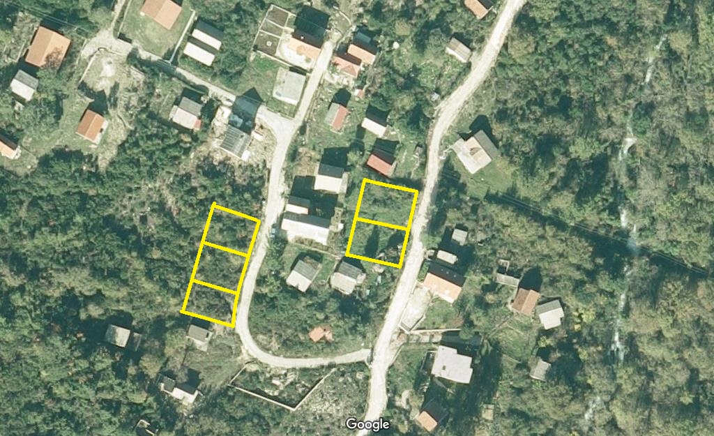
71387 Podlugovi-Ilijaš, BiH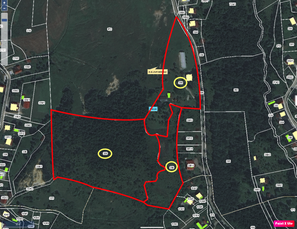 Prodaje se kuća sa pom. objektima i zemljištem: BREZA-SMREKOVICABreza, Breza, 71370, Bosnia and Herzegowina345,000 KM Prodaja
 Prodaje se kuća sa okućnicom: VISOKO - GORNJE MOŠTREGornje Moštre, Visoko, VISOKO, 71300, Bosnia and Herzegowina84,000 KM Prodaja
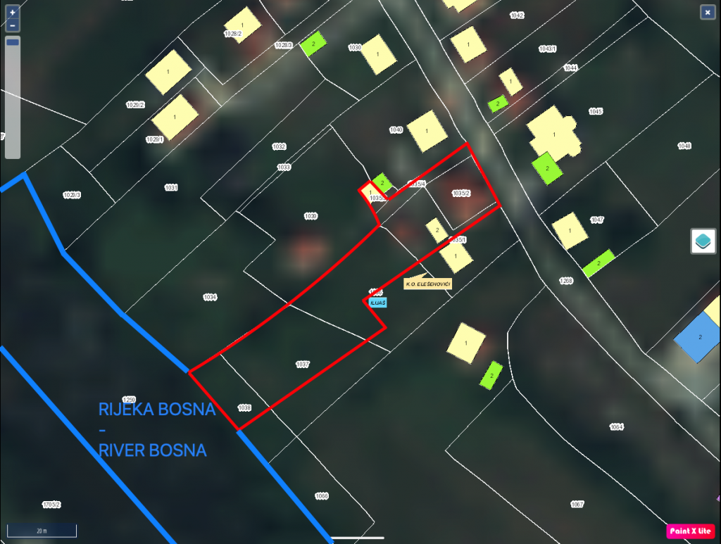 Prodaje se kuća s okućnicom: ILIJAŠ - LJEŠEVOIlijaš, Lješevo bb., Breza, 71380, Bosnia and Herzegowina293,000 KM Prodaja
 Prodaje se kuća sa zemljištem u Ilijašu - LJEŠEVOLješevo bb., Ilijaš, 71380, Bosnia and Herzegowina170,000 KM Prodaja
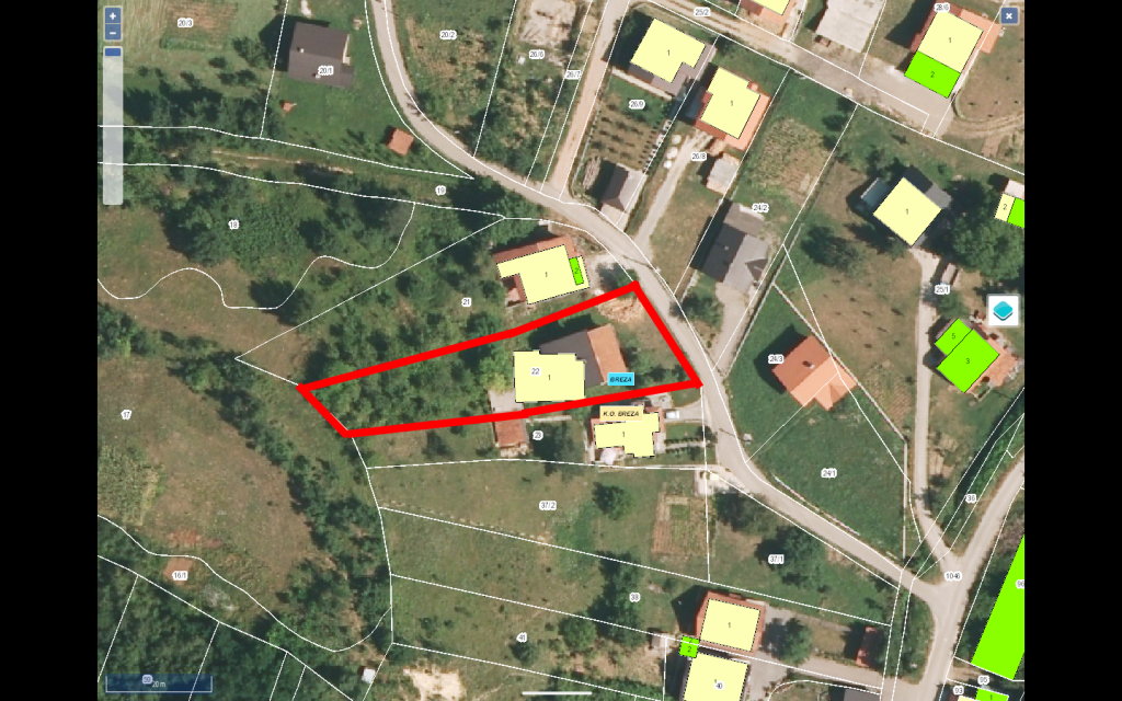 Prodaje se kuća sa zemljištem: Breza - STARE JAMEBREZA, BANJEVAC, BREZA, 71370, Bosnia and Herzegowina80,000 KM Prodaja
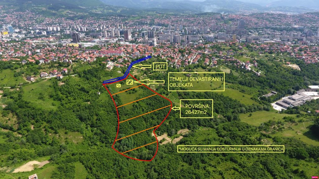 Prodaje se zemljište: SARAJEVO - Ul. HumskaHumska, Sarajevo, 71000, Bosnia and HerzegowinaKM Prodaja
Izdvojeno
Novo
- Prodaje se kuća sa pom. objektima i zemljištem: BREZA-SMREKOVICABreza, Breza, 71370, Bosnia and Herzegowina345,000 KM Prodaja
- Prodaje se kuća sa okućnicom: VISOKO - GORNJE MOŠTREGornje Moštre, Visoko, VISOKO, 71300, Bosnia and Herzegowina84,000 KM Prodaja
- Prodaje se kuća s okućnicom: ILIJAŠ - LJEŠEVOIlijaš, Lješevo bb., Breza, 71380, Bosnia and Herzegowina293,000 KM Prodaja